What is Phagocytosis and Pinocytosis?
Phagocytosis and pinocytosis are both important biological processes that help cells intake molecules or particles from outside into the cytoplasm. The main difference between these two processes is that phagocytosis helps intake solid particles while pinocytosis helps with small droplets of fluids.
Here, we will see what is phagocytosis and pinocytosis, in detail.
What is Phagocytosis?
The bulk transportation of large molecules, solid and foreign particles across the plasma membrane is called phagocytosis.
Phagocytosis occurs from outside the cell into the cytoplasm. It involves the continuous formation of invaginations to surround the target material, without any membrane extensions.
It is common in protozoans and eukaryotes. This process is usually seen in the granular leukocytes as well as in mesoblastic cells. The cells of mesoblastic origin are known as macrophagic or reticuloendothelial systems.
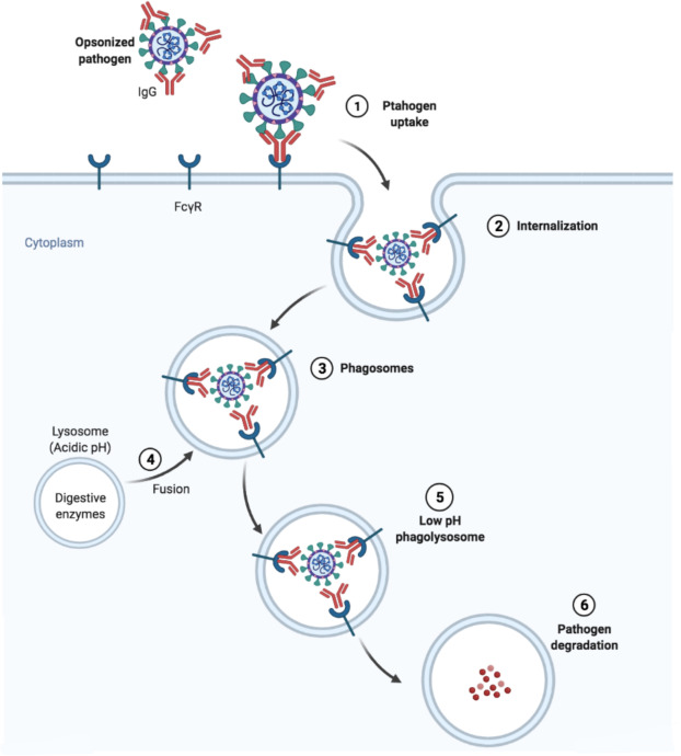
Process of Phagocytosis
Phagocytosis is a receptor-mediated mechanism. The mediators are called phagocytes such as DC, macrophages, monocytes, neutrophils, and mast cells.
- The target particles bind to the specific cell surface receptors in a process called adsorption.
- The plasma membrane expands its surface to engulf the adsorbed particle.
- The engulfed part becomes a vesicle called a phagosome.
- Phagosomes migrate into the cell and fuse with lysosomes to form the phagolysosome.
- Lysosome digests the target particle with the help of its digestive enzymes.
- The undigested food is expelled through plasma membrane in a process called egestion.
Types of Phagocytosis
Based on the physiology of the foreign particle, phagocytosis is classified into two types: ultra-phagocytosis and chromopexy.
- Ultraphagocytosis: Ultraphagocytosis is also called colloidopexy. It is the process of ingestion of smaller colloidal particles. It is mainly seen in macrophages and leukocytes.
- Chromopexy is the ingestion of chromogen particles.
Pinocytosis Definition
Pinocytosis is the non-specific uptake of small droplets of extracellular fluid with the help of pinosomes or endocytic vesicles, Materials are internalized in proportion to their concentration in extracellular fluid.
Edward observed pinocytosis in Amoeba and later by Lewis in cultured cells. Pino means drink and since the process is the intake of fluid, it is called pinocytosis.
Studies showed that Amoeba formed pinocytic channels on their cell surface through the invagination of plasma membranes. At the inner ends of each pinocytic channel, vesicles are pinched off to move toward the centre of the cell.
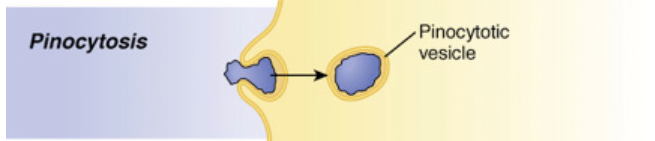
At the centre, these vesicles fuse with primary lysosomes to form food vacuoles. The contents are digested and the broken-down products are diffused into the cytoplasm.
Micropinocytosis
Micropinocytosis refers to pinocytosis occurring in submicroscopic levels of the cells.
Here, the invaginations of plasma membranes form smaller vesicles that are devoid of clathrin protein coat.
At the same time, the same process happens in cells of macrophages, reticular cells, ganglions, muscle cells, etc with the formation of clathrin-coated vesicles.
- Micropinocytosis is seen in endothelial cells of blood capillaries.
- Here, the vesicles detach from the plasma membrane and migrate to the plasma membrane on the opposite side.
- Upon reaching the membrane, it fuses with it and discharges the contents to expel it. This process is called transcytosis.
References
- Su, M., Parayath, N. N., & Amiji, M. M. (2017). Exosome-Mediated Communication in the Tumor Microenvironment. Diagnostic and Therapeutic Applications of Exosomes in Cancer, 187-218. https://doi.org/10.1016/B978-0-12-812774-2.00011-0
- Acharya, P. C., Fernandes, C., Mallik, S., Mishra, B., & Tekade, R. K. (2017). Physiologic Factors Related to Drug Absorption. Dosage Form Design Considerations, 117-147. https://doi.org/10.1016/B978-0-12-814423-7.00004-6
- Agarwal, P. V. |. V. (2004). Cell biology, Genetics, Molecular Biology, Evolution, and Ecology: Evolution and Ecology. S. Chand Publishing.
- Sadeghalvad, M., Mohammadi-Motlagh, H., & Rezaei, N. (2021). Structure and Function of the Immune System. Encyclopedia of Infection and Immunity, 24-38. https://doi.org/10.1016/B978-0-12-818731-9.00193-2
- Carlson, B. M. (2018). Cells. The Human Body, 1-25. https://doi.org/10.1016/B978-0-12-804254-0.00001-6


Hi! I know this is kind of off topic but I was wondering if you knew where I could find a captcha plugin for my comment form?
I’m using the same blog platform as yours and I’m having difficulty finding one?
Thanks a lot!
I like this website very much so much fantastic info .
excellent points altogether, you just gained a new reader. What would you recommend in regards to your post that you made a few days ago? Any positive?
After exploring a few of the blog articles on your site, I seriously like your technique of blogging. I book-marked it to my bookmark webpage list and will be checking back soon. Take a look at my website as well and let me know what you think. http://www.hairstylesvip.com
Greate article. Keep posting such kind of info on your site. Im really impressed by your site. Hi there, You have done an excellent job. I’ll definitely digg it and individually suggest to my friends. I am sure they will be benefited from this web site. http://www.kayswell.com