Describe the Structure and Function of Golgi Apparatus
Golgi apparatus was discovered by an Italian physician Camillo Golgi in 1898, in the nerve cells of barn owls and cats. It is present in all eukaryotic cells except a few cells such as mammalian RBC, sperm cells or bryophytes, and sieve tubes of plants. It is completely absent in prokaryotes.
The Golgi complex occupies different positions in different kinds of cells. In secretory and absorptive cells, it usually lies between the nucleus and cell surface where secretion or absorption occurs. In nerve cells, it surrounds the nucleus. In invertebrates and plant cells, the Golgi complex usually consists of many isolated units called dictyosomes scattered throughout the cytoplasm. Cytoplasm containing the Golgi complex has fewer or no other organelles and is called Golgi ground substance.
Structure of Golgi Bodies
Golgi bodies vary in size and form in different types of cells but they have a similar organisation. They appear as a coarse network under the light microscope. Electron microscopy shows it as a central stack or pile of parallel, flattened inter-communicating sacs or cisternae and many peripheral tubules and vesicles.
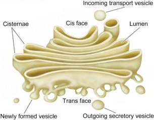
- The number of cisternae varies from 3-7 in most animal cells and 10-20 in plant cells.
- They look similar to smooth endoplasmic reticulum and are even continuous with them in some places.
- This suggests its origin from the smooth endoplasmic reticulum.
- Secretory materials reach the Golgi bodies through the vesicles that bud off from the smooth endoplasmic reticulum.
- They will fuse with the Golgi cisternae to form a face.
- Tubules are short.
- The anastomosing tubules arise from the periphery of cisternae with enlarged ends to form vesicles.
- Vesicles lie near the ends and the concave surface of the Golgi complex.
- They are pinched off from tubules of cisternae.
- They are of two types- smooth containing secretory molecules and the other is located that have an uncertain role.
- All Golgi bodies are filled with a fluid.
Chemical Composition of Golgi Bodies
Golgi bodies consist of a phospholipid bilayer sandwiched between two protein monolayers. Different enzymes originating from the smooth endoplasmic reticulum are associated with Golgi membranes.
Functions of Golgi Bodies
Formation of secretory vesicles
Golgi bodies process and package proteins and lipids transported from the endoplasmic reticulum for transport to other parts of the cell or outside of the cell. The packaging involves wrapping the materials in a membrane to form a secretory vesicle. Materials thus packed include zymogen in pancreatic cells, hormones from endocrine cells, etc. Secretory vesicles pinch off from the ends of cisternae and appear as dense secretory granules in the cytoplasm. Later, these vesicles release their contents by exocytosis.
Other functions of Golgi Bodies
- Synthesis of carbohydrates: It synthesises polysaccharides such as mucopolysaccharides, cellulose, pectic compounds etc from simple sugars.
- Formation of glycoproteins: Golgi bodies combine carbohydrates with proteins from the endoplasmic reticulum to form glycoproteins.
- Combines lipids and proteins from the endoplasmic reticulum to form lipoproteins.
- Add materials to the plasma membrane which were removed by endocytosis.
- Synthesises pectin and other carbohydrates necessary for the formation of cell walls.
- Produces mucilage and gums.
- Give rise to primary lysosomes by budding off from the maturing phase of cisternae.
- Acrosome formation in sperm cells
- Store secretions such as proteins and lipids.
- Vitellogenesis or formation of yolk in eggs.
- Formation of nematocysts and trichocysts.
- Absorption of materials.
References
- Agarwal, P. V. |. V. (2004). Cell biology, Genetics, Molecular Biology, Evolution, and Ecology: Evolution and Ecology. S. Chand Publishing.
- Cole, L., & Kramer, P. R. (2015). The Cell. Human Physiology, Biochemistry and Basic Medicine, 3-10. https://doi.org/10.1016/B978-0-12-803699-0.00001-3
Additional Reading
- Functions Of Cell Organelles
- Peroxisomes Structure and Function
- Glyoxysomes Structure and Function
- Structure and Function of Lysosomes


This blog post is worth the read – trust us!