Lysosomes are small membrane-bound vesicles with acidic lumen having hydrolytic enzymes. Here we will see the structure and function of lysosomes.
Explain the Structure and Function of Lysosomes
Lysosomes are associated with intracellular digestion or the digestion of different biological materials, ageing or death of cells, etc. They may also cause diseases such as cancer in animals.
Belgian cytologist and biochemist Christian De Duve discovered lysosomes in 1955. He called them perinuclear dense bodies. In 1956, Novikoff observed them under the electron microscope.
They occur practically in all animal cells including protozoans. They have also been found in certain plant cells such as yeast, fungi, root-tip cells of maize etc. Prokaryotes and bacteria lack lysosomes.
A few cell types such as mammalian RBC cells lack lysosomes. However, leukocytes are rich in lysosomes. It is also abundantly seen in absorptive, excretory and secretory organs as well.
Characteristics of Lysosomes
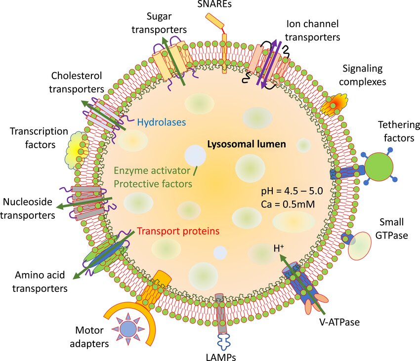
Lysosome lumen has a pH of around 5 which is amintianed through ATP-driven proton pumps across the cell membrane. More than 40 types of hydrolytic enzymes are seen in lysosomes, which include, proteases, nucleases, glycosidases, lipases, phospholipases, phosphatases, and sulfates.
- These are acid-dependent enzymes. It means they cannot perform their enzymatic activities in the normal pH of the cytoplasm thus protecting the other organelles in the cytoplasm.
- In addition, the membranes of lysosomes are resistant to these acidic enzymes thus preventing any possibility of a leakage.
- The materials needing hydrolysis must enter the lysosomes because these enzymes remain confined within them.
- In injured and dead cells, the lysosome membrane ruptures spontaneously releasing the enzymes that dissolve the weakened cells.
- In intact living cells, certain compounds such as cholesterol and cortisone prevent the rupturing of lysosome membranes.
- Cortisone, hydrocortisone etc stabilize the lysosomal membrane while lipo-soluble vitamins such as vitamins A, K, D, and E as well as sterols destabilize it.
Structure of Lysosomes
Lysosomes are generally spherical, vacuolar structures with a single membrane enclosing their dense material.
- Meristematic cells of plant roots have irregular lysosomes.
- A lysosome is a tiny sack bound by a single unit membrane of lipoprotein.
- It contains a dense, finely granular fluid. Later it will have digestive enzymes such as acid hydrolases.
- These enzymes can break down all major biological macromolecules in cells or those entering the cells from outside.
- The size and shape of lysosomes vary from one cell to another and also on their function.
- Their size varies from 0.2 to 0.8 mu.
- The lysosomal membrane is thicker than that of mitochondria.
- It has more carbohydrates, especially sialic acid.
- Lysosomal membrane proteins are highly glycosylated to protect from proteases inside.
- Lysosomal membrane fuses with other membranes in the cell.
- This membrane contains transport proteins that will release the final products of macromolecule digestion, back into cytosol for reuse or excretion.
Types of Lysosomes (Polymorphism)
Lysosomes are dynamic cell organelles showing polymorphism.
A. Animal Lysosomes
They are abundant in certain WBCs and secretory cells of the pancreas, spleen, liver, kidneys, and cells of degenerating or regressing tissue. Tadpoles’ tails contain many lysosomes.
Lysosomes change the nature of their contents at different times in the same cell on this basis 4 types of lysosomes are recognized. The first one is called primary lysosome while the other three are known as secondary lysosomes.
Primary Lysosomes
Primary lysosomes are also called proto lysosomes, storage granules or virgin lysosomes. They are newly formed having a diameter of 100 um. A newly formed one contains only degradative enzymes in an inactive state that do not participate in any processes. They become functional when they are converted into secondary lysosomes.
Heterophagosomes
When some material that is to be digested enters the primary lysosomes it is called a secondary lysosome digestive vacuole or heterophagosome. This commonly occurs by the fusion of a primary lysosome with a vacuole (pinosome or phagosome) or a secretory granule. These are also called heterophagic vacuoles.
The engulfed materials in the cytoplasmic vacuoles are digested by lysosome enzymes and are transported to the lumen. Here they become part of the matrix.
Autophagosomes
Autophagosomes are also called autophagic vacuoles, cytolysosomes, etc. A cell may digest its organelles like mitochondria and endoplasmic reticulum is called autophagy or autolysis.
The primary lysosome fuses around the damaged or unwanted organelles to form a large sack known as an autophagic vacuole, autophagosome or autolysosome. During cell growth or cell repair, the primary lysosomes digest intracellular structures like ribosomes, mitochondria, chloroplast, peroxisomes, etc through autophagy.
In some cases, the primary lysosomes engulf or enclose the cell organelle and later digest the outer membrane and then the inner membrane. Sometimes, the cell organelles are first enclosed by the smooth endoplasmic vesicles and then by lysosomes.
When the lysosomes engulf part of cytosol and digest it it is called microautophagy.
Residual Bodies
A secondary lysosome containing indigestible matter is called a residual body. They are also called dense bodies or telolysosomes which are often seen as large irregular bodies. They are a result of the incomplete digestion of food vacuoles.
In secondary lysosomes, the enzymes digest the incoming materials. These products of digestion pass through the lysosome membrane into the cytoplasm matrix to be used as a source of nutrition or energy. After digestion, the indigestible matter remains in the secondary lysosome.
The residual body meets the cell membrane and releases the residue by exocytosis. Residual bodies may also be stored in the cells which contributes to the ageing process.
This is due to the lack of some lysosomal enzymes. This undigested residue forms whorls of membrane, myelin figures, amorphous masses, etc. In Amoeba, they are eliminated by defecation while in others, they remain for a long time causing ageing.
B. Plant Lysosomes
In plants, lysosomes are not compartmentalized or confined to vacuoles. Many hydrolases are bound to the cell wall or in its vicinity.
They are not usually membrane-bound. Some storage granules with digestive enzymes are considered lysosomes. There are three kinds of plant lysosomes- vacuoles, spherosomes, and aleurone grains.
Vacuoles
Vacuoles are somewhat rounded sacs bound by a single-unit membrane. They are formed by the fusion of smaller vacuoles pro-vacuoles in the meristematic cells. They show little internal structure but contain a variety of hydrolytic enzymes. They are derived from endoplasmic reticulum and Golgi bodies with hydrolases. These enzymes are associated with tonoplast.
Spherosomes
Spherosomes are membrane-bound spherical bodies with granular contents rich in lipids and proteins. They occur in most plant cells and arise from the endoplasmic reticulum.
They originate from the endoplasmic reticulum where oil accumulates at the end and vesicles cut off to form pro-spherosomes.
Spherosomes are involved in lipid synthesis and storage. They are responsible for the digestion of cytoplasmic components by phagocytosis.
Aleurone Grains
Aleurone grains are membrane-bound spherical bodies containing proteins and phosphates. They are often associated with protein storage bodies such as in cells of endoplasm and cotyledons of seeds.
They are formed in the last stages of seed ripening and disappear in the early stages of germination. They also store phosphate as phytin.
Extracellular Digestion
Plant cells secrete hydrolases for extracellular digestion as seen in insectivorous plants producing proteases-rich liquid.
Functions of Lysosomes
- Digestion of useful materials- The organic substances taken up by cells in vacuoles from the environment are digested and this process is called intracellular digestion.
- Digestion of harmful materials- Foreign particles are disposed of by hydrolysing them in certain leukocytes and macrophages. This is called the natural defence of the body.
- Digestion of unwanted materials such as dead cells, debris etc that accumulate at the sites of injury are destroyed. This is natural scavenging of the body.
- Renewal of cells and organelles.
- Feeding of starving animals: Lysosomes provide food for starving animals by providing stored food materials and even the cells. This is called autophagy.
- Autolysis
- Helps in fertilization: Lysosomes of sperm cells release their enzymes to dissolve the egg membrane to facilitate the entry of the sperm nucleus during fertilization. This is called extracellular digestion.
- Lysosomes may be called the suicidal bags of the cell given their autolytic role or disposal units of the cell because they digest the incoming food materials and remove foreign bodies and the organelles that are no longer needed.
References
- Allemailem, Khaled & Almatroudi, Ahmad & Alrumaihi, Faris & Almatroodi, Saleh & Alkurbi, Mohammad & Basfar, Ghaiyda & Rahmani, Arshad & Khan, Amjad. (2021). Novel Approaches of Dysregulating Lysosome Functions in Cancer Cells by Specific Drugs and Its Nanoformulations: A Smart Approach of Modern Therapeutics. International Journal of Nanomedicine. 16. 5065-5098. 10.2147/IJN.S321343.
- Agarwal, P. V. |. V. (2004). Cell biology, Genetics, Molecular Biology, Evolution, and Ecology: Evolution and Ecology. S. Chand Publishing.
Additional Reading
- Characteristics of Cells
- Functions Of Cell Organelles
- Peroxisomes Structure and Function
- Glyoxysomes Structure and Function