NIOS Class 12 Biology Chapter 14 provides learners with solved terminal exercises that simplify exam preparation. The chapter includes important biology concepts explained in a clear and structured way.
These solutions help in quick revision and build strong conceptual understanding. By practising them, students can gain confidence and perform better in exams. This resource is a valuable part of NIOS Class 12 Biology studies, ensuring effective preparation for terminal and board examinations.
NIOS Class 12 Biology Chapter 14 Terminal Solutions
1. List the major steps that are involved with respiration in humans.
- (i) Breathing or pulmonary ventilation leading to the exchange of oxygen and carbon dioxide between the atmospheric air and the lungs.
- (ii) Exchange of gases at the alveolar surface.
- (iii) Transport and exchange of gases in the tissues.
- (iv) Cellular respiration.
2. How is oxygen transported in an earthworm?
Earthworm has no respiratory organs. The entire skin of the body of the earthworm functions as the respiratory surface.
- The skin of an earthworm is thin, moist, and has a rich supply of blood capillaries. Thus, it is very suitable for respiration.
- The body surface is covered with a moist film consisting of secretions of mucous glands, coelomic fluids, and excretory wastes.
- The capillaries on the skin take up O2 dissolved in the water (in the moisture) on the surface of the skin and release CO2 into the atmosphere.
- Earthworms have a closed circulatory system, which means that blood flows within blood vessels. The respiratory pigment haemoglobin remains dissolved in blood plasma and not in any cell.
- There is regular rhythmic contraction of blood vessels, which helps in the circulation of blood and hence in the transport of dissolved gases in the body.
3. Name the respiratory pigment in the earthworm.
Haemoglobin.
4. What is the role of carbonic anhydrase in the transport of carbon dioxide in our body?
Carbonic anhydrase acts as a catalyst to the reaction where CO2 and H2O combine to form carbonic acid (H2CO3) in the RBCs. The decomposition of carbonic acid produces hydrogen ions and HCO–3. This bicarbonate is easily soluble in blood plasma. It is carried to the alveoli and is released into the alveolar air to be expelled.
5. Which part of our respiratory system is known as the voice box?
The larynx is also known as the voice box. It is a small cartilaginous organ with vocal cords. It is also lined by ciliated epithelium. It connects the pharynx to the trachea and helps in sound production.
6. Where are the respiratory centres situated in our brain?
- (a) Dorsal respiratory group – generates basic respiratory rhythm. It stimulates the external intercostal muscles, the diaphragm contracts, and inspiration occurs. When the stimulation ceases, these muscles relax and expiration takes place.
- (b) Ventral respiratory group sends signals under enhanced respiratory needs. It controls both inspiration and expiration.
- (c) The Pneumotaxis centre in the pons controls the switch-off point of inspiration and thereby smoothens the transition between inspiration and expiration.
7. Name one nitrogenous waste removed by the kidney.
Uric acid.
8. Name the hormone the absence of which will result in excretion of hypotonic urine.
ADH (Anti Diuretic Hormone).
9. What is the role of cellophane in dialysis?
Cellphane tubes in artificial kidneys are permeable to micro molecules such as urea, uric acid, and mineral ions. It is not permeable to macromolecules such as plasma proteins.
10. Why is inspiration said to be an active phase and expiration a passive phase?
Inspiration
- A muscular dome-shaped diaphragm is present at the base of the lungs.
- On contraction, it becomes flattened and lowered.
- The lower surface of the lungs is pulled downwards, and the volume of the lungs increases.
- External intercostal muscles present between the ribs contract, the rib cage moves outwards and upwards.
- These contractions together increase the volume of the chest cavity, lower the air pressure within the lungs, and the atmospheric air rushes in, filling the lungs with fresh air.
- Thus, inspiration is an active phase of breathing.
Expiration
- Expiration (releasing air) involves the relaxation of external intercostal muscles and contraction of internal intercostal muscles.
- As a result, the rib cage lowers and moves inwards.
- The diaphragm also relaxes and rises again into its original dome-shaped condition.
- The abdominal organs press up against the diaphragm.
- This change decreases the volume of the chest cavity, thus increasing the air pressure within the lungs and the air, which is laden with CO2 and is forced out.
- 11. Differentiate between
- (a) Breathing and respiration
- (b) Inspiration and expiration
(a) Breathing and respiration
| Breathing | Respiration |
| Physical process | Biochemical process involving enzymes |
| Takes place only in reptiles, birds, and mammals | Occurs in all organisms |
| It is a rhythmic process | It is a continuous process |
| It is an extracellular process | It is an intracellular process |
| It involves gaseous exchange between the animal and its external environment | It involves the enzymatic breakdown of glucose in the presence or absence of oxygen to release energy. |
(b) Inspiration and expiration
Inspiration or taking air in, and expiration or forcing air out.
1. Inspiration (The intake of air):
- A muscular dome-shaped diaphragm is present at the base of the lungs.
- On contraction, it becomes flattened and lowered.
- The lower surface of the lungs is pulled downwards, and the volume of the lungs increases.
- External intercostal muscles present between the ribs contract, the rib cage moves outwards and upwards.
- These contractions together increase the volume of the chest cavity, lower the air pressure within the lungs, and the atmospheric air rushes in, filling the lungs with fresh air.
- Thus, inspiration is an active phase of breathing.
2. Expiration (releasing air):
- Expiration (releasing air) involves the relaxation of external intercostal muscles and contraction of internal intercostal muscles.
- As a result, the rib cage lowers and moves inwards.
- The diaphragm also relaxes and rises again into its original dome-shaped condition.
- The abdominal organs press up against the diaphragm.
- This change decreases the volume of the chest cavity, thus increasing the air pressure within the lungs and the air, which is laden with CO2 and is forced out.
12. List the special features of alveoli that enable easy gaseous exchange.
Alveoli are tiny sac-like structures. They have a thin and moist membrane rich with blood capillaries. Their wall has a single-layered flattened epithelium. It facilitates smooth gaseous exchange from the bronchioles.
13. What are vital capacity, tidal volume, and residual volume?
- Vital capacity is the volume of air that can be maximally breathed out after a maximum inspiration.
VC = IRV+TV+ERV)
- Tidal volume is the volume of air inhaled and exhaled without any noticeable effort (normal breathing).
- Residual volume is the volume of air that can be expelled by forced expiration over and above the normal expiration.
14. Give reasons for the following:
- (a) Exchange of gases at the alveolar surface continues even during expiration.
- (b) Trachea and bronchi do not collapse when air pressure decreases inside them.
(a) Exchange of gases at the alveolar surface continues even during expiration.
Even during expiration, there would still be some amount of air remaining in the lungs, which is called residual volume. This air is enough to continue gaseous exchange even during expiration.
(b) Trachea and bronchi do not collapse when air pressure decreases inside them.
The presence of cartilage, the C-shaped rings in the tracheal wall, protects the trachea and bronchi. The alveoli walls have Type-II cells that secrete surfactant that prevents them its collapsing during respiration.
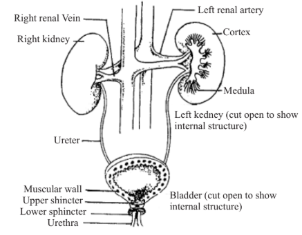
15. Draw the excretory system of the human and label the parts.
16. Draw the structure of a nephron and label the parts.
17. What are the causes and symptoms of pneumonia and TB?
Pneumonia
- Acute inflammation caused by diplococcus infection in the alveoli of the lung is the cause of pneumonia.
- It causes fever, pain, and a severe cough. Most of the air space is occupied by fluid and dead W.B.C.
TB- Tuberculosis
- TB or tuberculosis is a bacterial infection that spreads through droplets of an infected person.
- It can affect many other organs, but pulmonary TB is most common. Weight loss and cough are common symptoms. It is accompanied by a low fever. In extreme cases, blood may come out while coughing.
18. What is the role of the liver in excretion?
- It excretes bile pigments, cholesterol, drugs, and some vitamins.
- It excretes all the above-mentioned substances in bile, which flows into the small intestine, and from there these are removed with the faeces.
- Formation of urea and uric acid (from ammonia) also takes place in the liver. These are removed from the body by the kidneys.
19. Explain how nitrogenous wastes are removed from the body of a cockroach.
- The cells of Malpighian tubules remove nitrogenous waste and certain salts from the haemolymph and then pump them into the lumen of the tubule.
- Fluid passes to the hindgut and, in the process, gets concentrated.
- This concentrated fluid then moves into the rectum and is excreted as concentrated urine along with faeces.
- Most of the salt and water is pumped back into the haemolymph by Malpighian tubules, and in this way, the nitrogenous wastes are eliminated as almost dry matter.
20. How does ultrafiltration and reabsorption occur in nephrons?
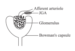
Each glomerular capillary receives blood flowing under high pressure through a branch of the renal artery. There is a continuous process of ultrafiltration (filtration under pressure).
All small molecules like water, glucose, minerals, amino acids, urea, and uric acid are filtered out of the blood plasma into the Bowman’s capsule through the capillary walls. Proteins remain in the glomerular blood. Thus, a protein-free filtrate is collected in the lumen of the Bowman’s capsule. The hydrostatic pressure of the circulating blood provides the filtration pressure.
As the glomerular filtrate flows through the tubules, several substances useful to the body, such as glucose and acids, and mineral ions needed to maintain the water and salt balance, are reabsorbed through the walls of the renal tubule.
21. Explain how gaseous exchange takes place in the lungs.
Air passes through the nostrils into the bronchi, to the bronchioles, and into air sacs, which are thin-walled sacs with a single layer of cells and heavily covered with blood capillaries. O2 from alveoli passes into capillaries, and CO2 from other capillaries diffuses into alveoli for removal. Alveoli are the organs where the actual gaseous exchange occurs.
22. How is oxygen transported from the lungs to the tissues and carbon dioxide from the tissues to the lungs?
Oxygenation of blood takes place in the lungs. Four molecules of oxygen form a reversible bond with haemoglobin, forming the compound oxyhaemoglobin.
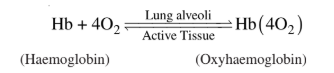
When the oxygenated blood reaches the tissue surface, there is a high concentration of CO2 in the tissues, oxygen having been used up, and a low concentration of O2.
As a result, the bonds holding oxygen and haemoglobin in Hb (4O2) become unstable and blood releases oxygen and takes up CO2.
Transport of carbon dioxide (from tissues to lungs)
Blood transports carbon dioxide with comparative ease because of its high solubility.
Active tissues constantly produce CO2. This CO2 is transported to the lungs in three ways:
- (i) CO2 is physically dissolved in blood plasma (only 5-7% of the total CO2 is transported).
- (ii) CO2 directly combines with haemoglobin of RBCs to form carbaminohemoglobin (about 21-23% only).
- (iii) As bicarbonate, it is dissolved in plasma but produced in RBCs, catalysed by the enzyme carbonic anhydrase, and then diffuses into plasma, the largest fraction of CO2, about 75% to 80%, to be transported in this manner.
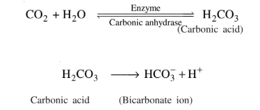
Bicarbonate is extremely soluble and dissolves in blood plasma. It again passes into the RBC and breaks down into CO2 and H2O in the alveoli. Inside the lungs, the CO2 is transported to the lungs from tissues in three ways mentioned above and is released into the alveolar air and finally breathed out.
23. How is (a)Water balance, and (b) Salt balance maintained by the kidney?
Water Balance
When the water content of the body is higher, leading to low osmotic pressure, less ADH (anti-diuretic hormone) is released. Hence, the wall of the DCT and collecting tubules remains less permeable, and as a result, plenty of dilute urine (hypotonic urine) is excreted.
When the water content of the body is low, the posterior pituitary secretes more of ADH. The permeability of the tubules is increased. As a result, more water is reabsorbed into the blood, and a reduced volume of concentrated urine is excreted (hypertonic urine).
Salt Balance
In response to low sodium ion concentration (or low blood pressure), another hormone, aldosterone, is released by the adrenal cortex. It stimulates the kidney tubules to absorb sodium ions in exchange for potassium ions. This leads to reabsorption of water by osmosis. As a result of increased blood volume, the blood pressure is increased. Similarly high sodium concentration will inhibit aldosterone release, and as a result, in would lead to a lower sodium ion concentration in the blood.
24. List the parts of the human respiratory system in correct sequence and state their functions.
| Organ | Function |
| Nostril | Filtration of unwanted particles. |
| Nasal Cavity | Traps dust, bacteria; warms and moistens the air in the pharynx. |
| Pharynx (throat) | The common passage for both respiratory gases and food moving into the digestive passage, separated by the epiglottis Epiglottis is a flap-like structure that closes the tracheal opening (opening of the wind pipe) called the glottis when food is swallowed. |
| Larynx (Voice box) | Connects the pharynx to the trachea; helps in sound production. |
| Trachea (Wind pipe) | Passage for air up to the bronchi. |
| Bronchus | Enters the lungs and divides to form secondary bronchi, tertiary bronchioles, and ultimately terminal bronchioles. Together they form thebronchial tree. |
| Bronchioles | Convey air into alveoli. |
| Alveoli (Air sacs) | Exchange of Gases. |
25. List three characteristics of our lungs that make them suitable as a respiratory surface.
- Lungs have thin-walled alveoli for gaseous exchange.
- Alveolar walls are highly permeable.
- There are numerous blood capillaries on the alveolar walls.