Mitochondria are specialized cell organelles in fungi, plants, and animal cells. Mitochondria are the powerhouse of the cell and power energy for the various cell functions. The word “Mitochondria” is derived from the Greek words- “mitos” meaning thread and “chondros” which means granule. Prokaryotes do not have mitochondria.
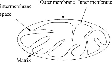
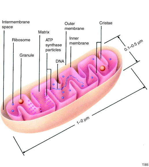
Structure of Mitochondria
Mitochondria have varying forms and sizes but are characteristic of each cell type. They are usually sausage-shaped but may be spherical, cylindrical, oval, filamentous, or branched as well. Spherical units are 1-5 mu in diameter.
For replication, mitochondria contain genetic information in the form of DNA, protein-making machinery such as ribosomes, and energy-producing mechanisms ( respiratory enzymes).
During cell division, each daughter cell receives some mitochondria from mother cells. These mitochondria later replicate to restore their normal number in the cell.
Mitochondrial Division
During mitosis, the number of mitochondria in the cell is equally divided and distributed to the daughter cells. This increase in their number occurs during the interphase. This mitochondrial division is similar to that of cell division.
- The inner membrane folds inwards and elongates.
- Some centrally located cristae produce partition foldings by the inner membrane.
- They fuse to form the separation between the matrix where they meet.
- Once the separation is formed, the outer membrane folds inwards meets each other, and separates the two mitochondria.
Biogenesis of Mitochondria
There are three hypotheses regarding the biogenesis of mitochondria.
- de Novo Hypothesis: According to the de Novo hypothesis, mitochondria are made of building blocks from lipids and amino acids. However, this was later rejected due to lack of evidence.
- Origin from Cell Organelles: It is postulated that mitochondria originated from the plasma membrane of cell organelles such as endoplasmic reticulum. This was also rejected due to lack of evidence.
- Origin from pre-existing Mitochondria: The most accepted hypothesis that is proven is the theory that mitochondria originated from pre-existing ones by their division. Each mitochondrion will double its mass and equally divide to form a new mitochondrion.
Function of Mitochondria
- Mitochondria are the main seat of cell respiration. They bring about stepwise oxidation of foodstuff or low-grade fuel for the cell and transfer the energy to release high-grade fuel, the ATP. This ATP is used to bring about energy-requiring activities in the cell. On this account, the mitochondria are often described as the powerhouse or storage batteries of the cell.
- Mitochondria provide intermediates for the synthesis of important biomolecules such as chlorophyll, cytological steroids, etc.
- Some amino acids are also formed in mitochondria.
Semi Autonomous Nature of Mitochondria
Mitochondria are capable of replicating itself and creating new mitochondrions from pre-existing ones. This is possible due to the presence of both DNA and ribosomes in it. They can produce the necessary proteins for their smooth functioning.
It shows the semi autonomous nature of mitochondria that do not completely depend on the nuclear DNA for a part of its protein requirement. However, most proteins for mitochondrial activities are made by the nuclear genes.
Since the proteins required by mitochondria are partially synthesized in the nucleus, mitochondria is not an independent or autonomous organelle.
Genomic Organisation of Mitochondria
Mitochondrial DNA is denoted as mtDNA. mtDNA molecules are smaller and simpler. It is double-stranded in all organisms except the protozoans and algae, where it is circular.
- mtDNA is larger in plants when compared to animals.
- It is located in the matrix.
- They are attached to the inner membrane where the DNA duplication starts.
- Duplication of mtDNA is controlled by the nuclear genes and enzymes in the cytosol.
- Mitochondrial ribosomes are called mitoribosomes or polyribosomes.
- These ribosomes are tightly associated with the mitochondrial inner membrane.
Mitochondrial Protein Synthesis
- Mitochondria synthesize around 12 different proteins.
- These proteins are incorporated into the inner membrane of mitochondria.
- Most of the proteins synthesized by mitochondria are hydrophobic.
- Chloramphenicol inhibits mitochondrial protein synthesis but does not affect protein synthesis in the cytosol.
- Cycloheximide does not affect mitochondrial protein synthesis but inhibits protein synthesis in the cytosol.
- The protein synthesis process in mitochondria is similar to that in the nucleus.
- Mitoribosome synthesizes,
- The three largest cytochrome oxidase units
- One subunit of Cytochrome b-c complex.
- 4 subunits of ATPase
- Some hydrophobic proteins.
Importing Mitochondrial Proteins
Mitochondria receives most of its proteins from the cytosol. These mitochondrial proteins have a specific transport mechanism. They pass through the mitochondrial membrane at specific contact sites.
Contact sites are the connecting points between outer and inner mitochondrial membranes. This process is facilitated by ATP hydrolysis and membrane gradient.
- Only those proteins with a signal peptide are allowed to pass.
- Once the protein passes through, the signal peptide is cut off.
- In case the protein also contains a hydrophobic signal peptide, there will be a second step where the hydrophobic signal peptide is unmarked when the first signal peptide is cut off.
- The unmarking helps the proteins to pass through.
- Once the protein is transported into the matrix, it is unfolded and ready to be used.
Lipid Biosynthesis in Mitochondria
Mitochondria synthesize lipids during their biogenesis or when producing new mitochondrion from existing ones. Otherwise, mitochondria imports most of their required lipids. These lipids are produced in the endoplasmic reticulum and are transported across the mitochondrial membrane.
- Phospholipid exchange proteins mediate this lipid transportation.
- The lipids cross the inner membrane at contact sites.
- Some of them undergo a decarboxylation reaction and are converted into cardiolipins.
- Cardiolipins constitute about 20% of total lipids.
References
- Agarwal, P. V. |. V. (2004). Cell biology, Genetics, Molecular Biology, Evolution, and Ecology: Evolution and Ecology. S. Chand Publishing.
- https://content.patnawomenscollege.in/zoology/Semi%20autonomus%20nature%20of%20mitochondria%201st%20year.pdf
- https://dacollege.org/uploads/stdmat/zoo-sem2-CC4-Unit3-Mitochondria-Structure-Semi-autonomous-nature.pdf
- Demirel, Y. (2001). Thermodynamics and biological systems. Nonequilibrium Thermodynamics, 293-355. https://doi.org/10.1016/B978-044450886-7/50012-6
Additional Reading
- Peroxisomes Structure and Function
- Glyoxysomes Structure and Function
- Structure and Function of Lysosomes
- Structure and Function of Golgi Apparatus