The network of proteins in the cytoplasm of a cell that maintains its shape keeps the organelles in their places, and helps vesicles navigate through the cytosol is called the cytoskeleton. Microtubules microfilaments and intermediate filaments form this cytoskeleton.
What are Microtubules?
Microtubules were first seen by Robert and Frankie in the axons of medullated nerve fibers. They called them neurotubules which were later found in the cytoplasmic matrix of all eukaryotes as well as in the cilia, flagella, sperm tail, etc. Microtubules are absent in amoeba and other prokaryotes.
Structure of Microtubules
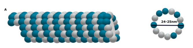
- Microtubules are hollow and unbranched cylinders usually about 200-270 A0 thick and several micrometers long.
- They have a diameter of 20-25 nm.
- They radiate from centrioles and extend to the periphery of the cells.
- The wall of a microtubule is composed of 13 parallel protofilaments that enclose a central lumen about 150 A0 wide.
- Each protofilament is made up of a protein called tubulin.
- A tubulin subunit contains one alpha-tubulin molecule and one beta-tubulin molecule.
Functions of Microtubules
- Microtubules form the cytoskeleton that maintains the shape and provides support to the cell and its processes.
- It forms motile elements of cilia and flagella to help in movements.
- Helps form the spindle apparatus during mitosis.
- Chromosomal fibers of the spindle bring about the movement of chromosomes to their opposite poles in anaphase.
- Cause movement of pigment granules in chromatophores thereby enabling their distribution.
- They are the components of centrioles and basal bodies.
- Serve as a track for the oriented transport of macromolecules in the cell, thus forming a microcirculatory system or intra-cellular transport.
Intermediate Filaments
Intermediate filaments are structural components that maintain the shape and structure of the cell. They do not take part in any functions of the cell. They are capable of bearing tensions and forming a structural scaffolding in the cell.
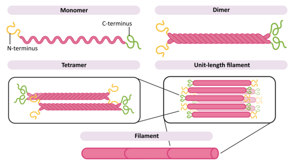
Intermediate filaments are made of fibrous proteins. The strands of protein wound together to form these filaments. Since these filaments have a size that falls between the microtubules and microfilaments, they are called intermediate filaments. They have a diameter of 10 nm.
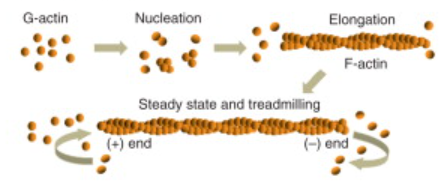
What are Microfilaments?
Microfilaments are found in all eukaryotic cells. They cause movements of microvilli to quicken the absorption of food. It is most prominent in the muscle cells and are called myofilaments. They are absent in prokaryotic cells.
They are solid, unbranched, rod-like fibrils of indefinite length and about 3-6 nm in diameter.
Microfilaments are composed of a globular protein called G-actin and the filamentous ones are made of myosin.
Functions of Microfilaments
- Forms a cytoskeleton to support the relatively fluid matrix.
- It helps in the directed movements of particles and organelles in the cell.
- Enable streaming movement of cytoplasm during interphase between stationary ectoplasm and moving endoplasm.
- Creates cleavage or furrow during cell division.
- Participate in the gliding amoeboid motion, and movements of leukocytes and macrophages.
- Responsible for change in the cell shape during development, motility, and division.
- Contraction of muscles.
- Movement of villi to quicken absorption of food.
- Movement of the plasma membrane during endocytosis.
References
- Agarwal, P. V. |. V. (2004). Cell biology, Genetics, Molecular Biology, Evolution, and Ecology: Evolution and Ecology. S. Chand Publishing.
- https://www.kharagpurcollege.ac.in/studyMaterial/2759SEM-2-Cytoskeleton-Zoology-Lecture-1-for-sem-2-04-04-2020.pdf
- https://bio.libretexts.org/Bookshelves/Introductory_and_General_Biology/General_Biology_1e_(OpenStax)/2%3A_The_Cell/04%3A_Cell_Structure/4.5%3A_The_Cytoskeleton
- Frixione, E., & Hernández, M. (2010). Structural Organization of Cells – The Cytoskeleton. Comprehensive Biotechnology (Second Edition), 367-381. https://doi.org/10.1016/B978-0-08-088504-9.00039-8