The endoplasmic reticulum (ER) is the complex network of membrane-bound interconnecting vacuoles or cavities in the cytoplasm. It was discovered by Porter in 1945 and was named in 1953.
The name endoplasmic reticulum was derived from ‘reticules’, the netted purse that women carry. Due to its likeness of endoplasmic reticulum to the net, embedded in the cytoplasm, it was named so.
Characteristics of Endoplasmic Reticulum in Plant Cell
- ER is common in all cells except egg cells, embryonic cells, and erythrocytes (RBC).
- It is poorly developed in immature RBCs.
- In some cells, only smooth endoplasmic reticulum is seen while some cells have only rough endoplasmic reticulum.
- Liver cells contain both rough and smooth endoplasmic reticulum.
Endomembrane System
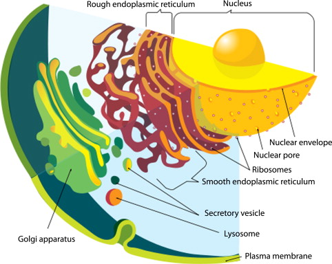
ER is a major part of the endomembrane system. This system encompasses the nuclear envelope, endoplasmic reticulum, and the Golgi apparatus. Each of these organelles of the endomembrane system is membraned. These membranes have a cytoplasmic and luminal face.
Morphology of Enodplasmic Reticulum
Morphologically endoplasmic reticulum is composed of the following three kinds of structures- cisternae, vesicles, and tubules.
Cisternae
- Cisternae are long and flattened, sac-like unbranched and tube-like structures.
- They have a diameter of 40-50 mu and remain arranged parallel in stacks.
- Rough endoplasmic reticulum exists as cisternae.
- They are usually seen in those cells that have synthetic roles as the cells of the pancreas, notochord, brain, etc.
Vesicles
- Vesicles are membrane-bound oval-shaped vacuolar structures having a diameter of 25-30 mu.
- They often remain isolated in the cytoplasm and occur in most cells but are specifically abundant in pancreatic cells.
- Vesicles are abundant in smooth endoplasmic reticulum.
Tubules
- Tubules are branched structures forming a reticular system along with cisternae and vesicles.
- They usually have a diameter from 50-190 mu and occur in almost all cells.
- The tubular form seen in the smooth endoplasmic reticulum is dynamic.
- They take part in membrane movements, fusion, and fission between membranes.
Ultrastructure of Endoplasmic Reticulum
- The cavities of cisternae, vesicles, and tubules of the endoplasmic reticulum are bound by a thin membrane of 50-60 A thickness.
- The membrane of the endoplasmic reticulum is trilaminar and has a fluid mosaic configuration, like a unit membrane, the plasma membrane, the nucleus, the Golgi complex, etc.
- The membrane is thus composed of a bilayer of phospholipid in which proteins of various sorts “float”.
- The membrane remains continuous with the plasma membrane, nuclear membrane and Golgi complex.
- The cavity of the endoplasmic reticulum is well developed and acts as a passage for secretory granules in the cavity of ER.
Types of Endoplasmic Reticulum
There are two types of endoplasmic reticulum- rough endoplasmic reticulum and smooth endoplasmic reticulum.
Granular or Rough Endoplasmic Reticulum
The type of endoplasmic reticulum having ribosomes attached to its membrane is known as granular endoplasmic reticulum or rough endoplasmic reticulum. These ribosomes play an important role in protein synthesis.
RER is commonly found in pancreatic cells, plasma cells, goblet cells, endocrine gland cells, and liver cells.
Due to the RNA content in ribosomes, RER takes in basophilic stains. The region of the matrix having RER takes in basophilic stain and is called ergastoplasm, basophilic bodies, chromophilic substances, or nissl bodies, etc by early cytologists.
Annulate Lamellae
Generally, the endoplasmic reticulum lacks pores. However, in some cases as in invertebrates, spermatocytes, ovocytes of vertebrates, etc, pores may be seen. These pores are formed by the invagination of the nuclear envelope. These invaginations form a stacked-sheet structures. These structures are called annulate lamellae which are continuous with the ER membrane.
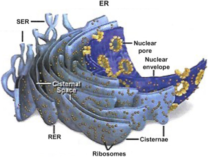
Agranular or Smooth Endoplasmic Reticulum
The type of endoplasmic reticulum having no ribosomes on its membrane will have a smooth surface and is thus known as smooth endoplasmic reticulum or agranular endoplasmic reticulum.
- SER is seen only in those cells that have no active participation in protein synthesis.
- Cells that are concerned largely with lipid synthesis have well-developed SER, especially those that metabolize glycogen and lipids.
- Eg, Cells of the adrenal cortex (which synthesizes steroid hormones) and intestinal absorptive cells have abundant SER.
- It is also seen in adipose cells, glycogen-storing cells of the liver, spermatocytes, and leukocytes.
- Muscle cells are also rich in SER which is called sarcoplasmic reticulum.
- In pigmented retinal cells, they exist in the form of tightly packed vesicles and tubules called myeloid bodies.
- Glycosomes are dense particles of fine tubules seen in glycogen-rich regions of the liver cells. They contain enzymes for the synthesis and conduction of fiber of the heart.
Enzymes of Endoplasmic Reticulum Membranes
The membranes of the endoplasmic reticulum contain several important enzymes for synthetic activities. The most important enzymes are stearases, NADH, cytochrome C reductase, NADH diaphorase, glucose-6-phosphatase, and Mg++-activated ATPase.
Certain enzymes of the endoplasmic reticulum such as nucleotide diphosphate, ascorbic acid, glucuronide, steroids, and hexose metabolism.
| Enzymes | Location |
| Mostly on cytoplasmic, also on the luminal face | Cytoplasmic face |
| NADH- Cytochrome b5 reductase | Cytoplasmic face |
| NADP- Cytochrome c reductase | Cytoplasmic face |
| Cytochrome P-450 | Mostly on cytoplasmic, also on luminal face |
| ATPase | Cytoplasmic face |
| 5 ́-nucleotidase | Cytoplasmic face |
| Nucleoside pyrophosphatase | Cytoplasmic face |
| GDP-mannosyl transferase | Cytoplasmic face |
| Nucleoside diphosphatase | Luminal face |
| Glucose -6- phosphatase (histochemical marker enzyme) | Luminal face |
| Acetanilide-hydrolysing esterase | Luminal face |
| β- glucuronidase | Luminal face |
The enzymes of the endoplasmic reticulum perform the following important functions
- Synthesis of glycerides such as triglycerides, phospholipids, glycerol, and plasmalogens.
- Metabolism of plasmalogens.
- Synthesis of fatty acids
- Biosynthesis of steroids like cholesterol, steroids, hydrogenation of unsaturated bonds.
- Oxidation of NADPH2 requires steroid transformations, aromatization, and aromatic hydroxylation, side chain oxidation, deamination, thioether oxidation, desulfuration.
- L-ascorbic acid synthesis.
- UDP-Uronic acid metabolism
- UDP-glucose dephosphorylation
- Aryl and steroid sulfates
Functions of Endoplasmic Reticulum
The endoplasmic reticulum acts as a secretory, storage, and circulatory system for the cell. While some functions are common to both SER and RER, there also are some functions that are exclusive to either SER or RER.
Common Functions of SER and RER
- They provide a structural framework to the cells and give mechanical support to the colloidal cytoplasmic matrix.
- It enables the exchange of molecules by a process of osmosis, diffusion, and active transportation through the membrane of the endoplasmic reticulum.
- ER has carriers and permeases that enable this transport across the membrane.
- Both SER and RER contain various enzymes for metabolic activities.
- It increases the surface area for enzymatic reactions.
- They serve as an intracellular circulatory and transport system for secretory products synthesized in RER and are transported through the SER, Golgi bodies, and lysosomes.
- Facilitates membrane flow of RNA and nucleoproteins from the nucleus to the cytoplasm.
- Conducts intracellular impulses from the surface membrane to deep muscle fibers.
- ER forms the new nuclear envelope after a nuclear division.
- The sarcoplasmic reticulum releases calcium when the muscle is stimulated and also takes it back to relax the muscles.
Functions of Smooth Endoplasmic Reticulum
The main functions of SER are lipid synthesis, glycogenolysis, sterol metabolism, and detoxification, among others.
Lipid Synthesis
Lipids such as cholesterol, phospholipids, etc are synthesized in the SER. The newly synthesized lipids are transported to other membranes by phospholipid exchange proteins.
Glycogenolysis
Glycogenolysis is the breakdown of glycogen with the help of glucose-6-phosphatase which is present in the SER membrane. It happens in the cytosol of glycosomes with the help of UDPG-glycogen transferase. This enzyme adds uridine diphosphate glucose to the primer glycogen and catalyzes the release of glucose in the SER lumen from the glucose-6-phosphate.
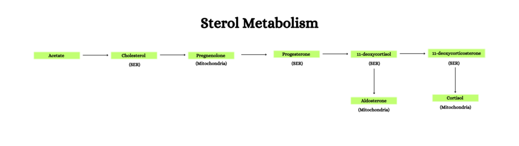
Sterol Metabolism
SER synthesizes cholesterol with its enzymes. Cholesterol is the precursor for bile acids and steroid hormones.
- Biosynthesis of cholesterol happens in 20 steps from acetate. 11 enzymes, that are bound to the SER, are required to these steps. Eg. Squalene synthase and HMG-Co A reductase.
- Cholesterol is converted into bile acids with the help of the cholesterol 7 alpha-hydroxylase enzyme.
- It also requires NADPH and oxygen molecules which are dependent on cytochrome P-450 and NADPH-cytochrome-c reductase from the electron transport chain of SER.
- Enzymes required for the biosynthesis of steroids and hormones in various organs are also located on the SER.
Detoxification
ER modifies toxic substances to become hydrophilic to help with their excretion. These toxic substances include drugs, insecticides, pollutants, carcinogens, etc. The enzymes required for the detoxification of these substances come from the RER first and then from the SER.
Other Functions of SER
SER is part of the synthesis of triglycerides in the intestine, visual pigments in the retina, and the formation of cellulose in the cell wall of plant cells.
Functions of Rough Endoplasmic Reticulum
The main function of RER is protein synthesis. Membrane proteins, proteins that are secreted and need to be exported are synthesized by the ribosomes on the RER.
RER provides the surface area for the process. These ribosomes are attached to receptors or specific binding sites by their 60s subunit. The synthesized proteins will pass from ribosomes into the cisternae of the RER to protect them from protease enzymes in the cytoplasm.
This transport happens during translation. The polypeptide chain upon reaching cisternae folds into its scion and testi structure.
Protein Glycosylation
The addition of sugar to secretory proteins is another function of RER. This process occurs in its lumen. The oligosaccharides are attached to the NH2 group of asparagine residue by the glycosyltransferase on the ER membrane.
References
- Agarwal, P. V. |. V. (2004). Cell biology, Genetics, Molecular Biology, Evolution, and Ecology: Evolution and Ecology. S. Chand Publishing.
- Litwack, G. (2017). The Cell. Human Biochemistry, 19-37. https://doi.org/10.1016/B978-0-12-383864-3.00002-8
- Stillwell, W. (2012). Introduction to Biological Membranes. An Introduction to Biological Membranes, 1-12. https://doi.org/10.1016/B978-0-444-52153-8.00001-5
Additional Reading
- Structure and Function of Golgi Apparatus
- Types of Plastids and Their Functions
- Ribosomes: Structure and Function