Mitosis is a crucial process in the cell cycle where the parent cell divides to produce two identical daughter cells with the same number of chromosomes. This is ensured by distributing the replicated DNA equally between the two daughter cells.
This cell duplication or division is an essential phase of the cell cycle that helps the organism with its repair, growth, and asexual mode of reproduction. Mitosis proceeds through various stages from preparing to completing the separation of the new cells formed.
Different Stages of Mitosis
The different stages of mitosis are,
- Prophase
- Prometaphase
- Metaphase
- Anaphase
- Telophase
1. Prophase
Prophase is the longest stage of mitosis and lasts from 1 to several hours. The chromosomes that remain in a diffused stage become more and more distinct as the cell enters prophase. This stage includes all the stages that transform the chromonemata from the metabolic stage into compact, separate individual bodies – the chromosomes.
The chromosomes become increasingly stainable as the prophase advances. This is due to three processes: dehydration, nucleation and spiralization. It is observed that there is a progressive reduction in the water content of chromosomes. The reduction of water content enhances the stainability of chromosomes.
Another factor that contributes to the increasing stainability of chromosomes is that the nucleic acid synthesised during the interphase gets organised on the chromosomes, and this process is called nucleation. The chromosomes become progressively thicker, shorter and increase in diameter. This process is called spiralization.
Each prophase chromosome is composed of 2 coiled identical filaments called sister chromatids. They are held together by a centromere. The two chromatids are closely associated along their length but do not fuse.
During early prophase, the chromosomes are evenly distributed in the nuclear cavity, but as prophase progresses, the chromosomes start approaching the nuclear membrane. This movement indicates the disintegration of the nuclear membrane.
- In prophase, the threadlike chromatin fibres condense around the histone proteins and become more compact.
- The reticulate nature of chromonemata becomes uniform, and threads belonging to different chromosomes stand out clearly.
- Chromosomes become shorter in length and increase in thickness.
- DNA become more visible and the enlargement of the nucleus occurs by the intake of water.
- The nucleolus gradually disappears, and the components of both the nuclear pore complexes and the lamin fibres become hyperphosphorylated.
By the end of the prophase, the fragmentation of the nuclear envelope is complete. The nucleolus also disperses and gradually disappears. The most conspicuous cytoplasmic change during mitotic prophase ( late ) is the formation of a spindle, which is made up of microtubules.
The appearance of spindles is related to the existence of centrioles in animal cells. Plant cells lack centrioles but instead possess polar caps, which take part in the cell division. The spindle mechanism helps the regular separation of daughter chromosomes to opposite poles.
2. Prometaphase
While the disappearance of the nuclear envelope marks the end of prophase, it marks the start of the prometaphase stage. The disintegrated nuclear envelope forms small vesicles and gets distributed between the daughter cells.
- Disintegration of the nuclear envelope gives the centromeres and microtubules access to the chromosomes.
- The microtubules get in contact with the chromosomes, and the latter attach to them at the chromosome kinetochores- the complex, platelike structures.
- As the prometaphase progresses, the attached chromosomes are tugged towards the opposite poles by the microtubules.
- The sister chromatids do not separate at this stage.
- As this stage comes to an end, the bivalent chromosomes are seen attached to the microtubules, ready to be pulled towards the poles.
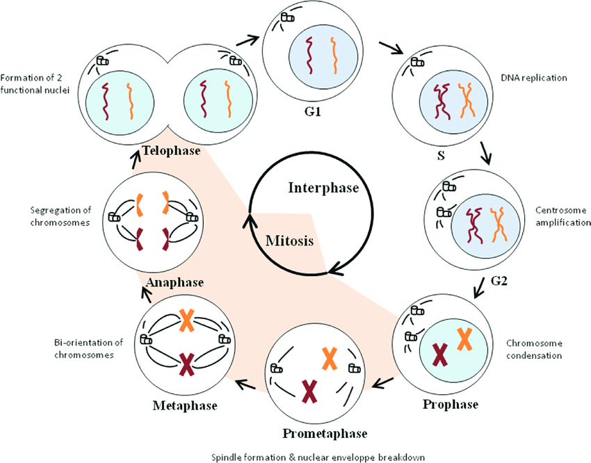
3. Metaphase
In the cell division cycle, the metaphase lasts only for a short duration. The metaphase chromosomes consist of two chromatids lying side-by-side and attached to the spindle by the centromere.
The fibres of the spindle that carry the chromosomes are called chromosomal fibres. The fibres that are seen in between the separating daughter chromosomes are called interzonal fibres, and those that extend without any interruption from one pole to the other are called continuous fibres or polar fibres. The spindle apparatus is made up largely of protein ( microtubules ) and has a comparatively lesser amount of RNA.
- During metaphase, the microtubules extend between the poles.
- The freely distributed condensed chromosome moves to the equatorial plate oriented along the central line.
- The movement of the chromosomes to the equator is called congressional.
- The chromosomes orient on the equatorial plate such that the centromeres are arranged on the longitudinal axis of the spindle, facing the poles and the arms lying freely in the cytoplasm.
- The chromosomes are irregularly scattered on the equatorial plate ( in plant cells ).
- The smaller chromosomes lie at the centre while the larger ones are at the periphery.
- Towards the end of metaphase centromeres of each chromosome divide longitudinally.
The assembly of chromosomes in this manner is crucial, as only those cells that have the correctly assembled spindle can enter anaphase. On the other hand, cells can be arrested at this stage, metaphase, when exposed to colchicine, which is known as a mitotic poison.
4. Anaphase
The process of mitosis is manifested in its climax at anaphase. Anaphase is initiated with the division of the centromere at the late metaphase. Soon after the division of the centromere, the daughter chromatids, which are now called daughter chromosomes, move toward the opposite poles. The movement of chromosomes in dividing cells follows certain regularities:
- In this stage, the kinetochore shortens, and chromosomes tend to move in a straight line.
- Movement proceeds at a decreasing speed.
- All chromosomes begin their movement at the same time and follow the same speed.
- The movement begins only after the centromeres of all chromosomes have divided.
- As the anaphase progresses, the distance between the chromosomes and the poles decreases while the distance between each chromosome increases on their fibers.
The movement of chromosomes to opposite poles can be explained in many ways. Some cytologists hold the opinion that it is affected by the stretching of the interzonal fibres, while some others believe that the contraction of chromosomal fibres brings the chromosomes to the poles. There are still others who advocate the repulsive force existing between the daughter centromeres as an effective force to pull the daughter chromosomes apart.
One of the accepted views is that the energy required for the movement of chromosomes is derived from DNA, which reacts with the protein of the spindle to liberate energy.
5. Telophase
The end of polar migration of the two daughter groups of chromosomes marks the beginning of telophase. Here, the interphase nucelus is restored gradually.
- The chromosomes start to uncoil and disappear.
- Proteins of the inner nuclear envelope associate with the chromosomes as pieces of the nuclear envelope rebind to the condensed chromosomes.
- These nuclear envelope pieces come in contact with each other and fuse to form the new nuclear envelope, or the nuclear membrane.
- The nuclear pore complexes reassemble into this envelope, connecting the nucleoplasm and cytoplasm.
- The nucleolus also condenses and reappears.
- At this time, the chromatin decondensation also happens.
- Thus, two daughter nuclei are formed at the end of karyokinesis.
6. Cytokinesis
Karyokinesis is followed by the division of cytoplasm; the failure of which results in a coenocytic condition. The division of cytoplasm (cytokinesis) in plant cells takes place with the formation of a cell plate, usually at the equatorial region of the spindle.
- A collection of vesicles called phragmoplast accumulates around the microtubules of the midplate of the spindle after the chromosomes have been pulled away to the poles.
- The vesicles are derived from the Golgi bodies.
- They fuse to form a disc or cell plate, which then grows outward and ultimately reaches the cell walls on either side of the cell.
- The cell wall grows and matures with the cells.
- In animal cells, the division is affected by furrowing.
Significance of Mitosis
Mitosis is significant for many reasons, as it plays an important role in the growth and reproductive processes of an organism.
- Mitosis is an extremely regular process. It maintains the qualitative and quantitative distribution of hereditary materials to daughter cells.
- It maintains the constancy of the diploid number of chromosomes of a species.
- It maintains the constancy and balance in the DNA and RNA content of the cell.
- Mitosis maintains the nuclear and cytoplasmic balance in the cell. The cells cannot expand indefinitely, it has to maintain a balance between the cytoplasm and nucleus called the nucleoplasmic index.
- Mitosis helps the cells maintain their proper size.
- It results in the growth and development of the organism
- Repair and replacement of dead cells is ensured by mitosis.
- It plays an important role in asexual reproduction in the formation of gametes.
- Mitosis is of great significance in cancer because it is the result of uncontrolled division. There are many mitotic abnormalities resulting in hazardous growth.
References
- Agarwal, P. V. |. V. (2004). Cell biology, Genetics, Molecular Biology, Evolution, and Ecology: Evolution and Ecology. S. Chand Publishing.
- McIntosh, J. R. (2016). Mitosis. Cold Spring Harbor Perspectives in Biology, 8(9), a023218. https://doi.org/10.1101/cshperspect.a023218
- O’Connor, C. (2008). Cell Division: Stages of Mitosis. Nature Education 1(1):188
Additional Reading
- Short Note on Chromosome
- Describe The Structure Of Chromosome
- Write a Short Note on Cell Division
- Explain The Mitotic Cell Cycle
Hey there outstanding blog! Does running a blog such
as this take a massive amount work? I’ve absolutely no understanding of programming however I had been hoping to start my own blog soon. Anyhow, should you have any recommendations or tips for new blog owners
please share. I understand this is off subject but I simply had
to ask. Thank you!
Great work! This is the kind of information that should be shared around the web. Shame on the search engines for now not positioning this put up higher! Come on over and consult with my site . Thanks =)