Chromosomes, the thread-like structure that carries the DNA in the nucleus is not visible in general. It appears only during cell division. When it does, it will have a distinct structure with various parts.
Here is how to describe the structure of chromosomes.
Structural Composition of Chromosome
The composing material of chromosomes is chromatin which is formed of DNA and nucleoproteins, which can exist as euchromatin and heterochromatin regions.
- Heterochromatin is the region that remains condensed during interphase and prophase.
- The non-condensed regions are called euchromatin.
- Due to these areas, chromatin has differential staining properties and this is called heteropyknosis.
- Heterochromatin and euchromatin are seen with the electron microscope and differ in their physical structure.
- Heterochromatin represents a condensed inter-coiled state with 2-3 times more chromatin than euchromatin.
- This region is transcriptionally inactive during mitosis.
- However, the euchromatin is less condensed and remains in its active state.
| Euchromatin | Heterochromatin |
| Chromatin fibers loosely coiled | Chromatin fibers tightly packed |
| Deeply stained in divisional cycle | Chromatin fibers are tightly packed |
| Less stained in interphase | Deeply stained in interphase |
| Phenotype affected due to loss or addition in this region | Phenotype unaffected by loss or addition in the region |
| Can synthesize mRNA invitro | Cannot synthesize mRNA invitro |
| Has a higher crossover frequency | Has low crossover frequency |
| Shows heteropycnosis | Does not show heteropycnosis |
| Less affected by age, sex, temperature, etc. | More affected by age, sex, temperature, centromere position etc. |
Chemical Composition of Chromosomes
- Nucleic acids and proteins are the most important chemical constituents of chromosomes. Quantitatively chromosomes contain 90% Deoxyribo-nucleoproteins and just 10% residual chromosomes.
- The former part contains 45% DNA and 55% histoproteins.
- The residual chromosomes contain 12-14% RNA, and 23% DNA and the remaining contain residual protein.
- The residual protein is an acidic protein containing large amounts of amino acids, tryptophan, and tyrosine.
- It is highly essential for the chromosome as its removal can affect its integrity.
- The removal of DNA or histone proteins will not affect the structure of the chromosome.
- The protein component serves as the framework to which the nucleic acids are attached.
Morphology of Chromosomes
The morphology of chromosomes such as the size and shape, can be studied from microscopic examination. It will stay constant for a particular species.
- Organisms having a large number of chromosomes may still be small in size and those having fewer chromosomes may be larger.
- In the cotton plant, Gossypium hirsutum, there are 52 chromosomes and Solanum tubersoum and Nicotiana tobacum have 48 chromosomes each.
- Sugar cane has 80, rice has 84, onion 16, and so on.
- Chlamydomonas has 10 chromosomes while bread mold carries just 2 chromosomes.
Size of Chromosomes
The size of chromosomes is measured in the metaphase and anaphase when they get thicker. It varies with species but stays relatively constant for a particular species.
- The length of chromosomes can be 0.2 to 50μ in diameter and 6μ in length.
- Monocots have larger chromosomes than dicots.
- Plants in general have larger-sized chromosomes than animals.
- The size of chromosomes inside the cell may vary from cell to cell.
- The largest chromosomes are the Lampbrush chromosomes found in vertebrate oocytes and polytene chromosomes in Dipteran insects.
Number of Chromosomes
Usually, two terms- haploid and diploid, are used to designate the chromosome number of an individual organism. Gametic cells such as sperms and ova usually have a single set of chromosomes and are termed haploid. It is also known as the genome.
On the other hand, the somatic cells or body cells of an organism usually have two sets of chromosomes and are called diploid chromosomes. Cells that carry such chromosomes are diploid. These diploid cells are a result of the union of two haploid cells, or gametes via sexual reproduction.
The entire collection of chromosomes in the nucleus of an individual is termed chromosome complement or karyotype. In a diploid chromosome complement, the two members of a pair of similar chromosomes are called homologous chromosomes.
Shape of Chromosomes
The shape of chromosomes changes with the phase of cell division and as part of the continual cell growth. During the resting phase, the chromosome appears as a thin, elastic, coiled, contractile thread that can be stained. These are called chromatin threads.
The shape of the chromosome is determined by the primary constriction which is located at the point where the arms or chromatids meet. Within the constriction, there is a clear zone containing a granule or spherule. This is called centromere or kinetochore.
- Chromosomes of most organisms contain only one centromere and are called monocentric chromosomes.
- Certain chromosomes may have two or more centromeres and are thus called dicentric or polycentric chromosomes.
- In some cases, centromeres are found all along the chromosomes and are called diffused chromosomes.
- If the centromere is absent, this chromosome will be acentric. During cell division, the centromere may lag and can get lost.
Structure of Chromosomes
According to Lima De Faria, the structure of the centromere may be more complex. During cell division, the middle zone maintains the chromosome’s connection to the spindle. In the metaphase when the sister chromatids form, they will still be connected by a region that has a special division cycle.
Besides the primary constrictions, a chromosome must also have a few secondary constrictions that are different from the other one due to the absence of angular deviation of chromosomic segments. Some of these secondary constrictions are important for the formation of nucleolus and are called nuclear zones or nucleolar organizers. Generally, only two chromosomes of a nucleus have these nucleolar zones and they are called nucleolar chromosomes.
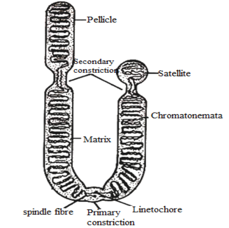
- The matrix will be a viscous liquid which is limited from the outside by a membrane called a pellicle.
- The matrix and pellicle are made of non-genetic materials.
- This early concept that the chromosomes consist of the limiting membrane and the amorphous matrix between the coils of chromonema is now rejected altogether after observations under the electron microscope.
- Each chromatid is formed of two thin longitudinal chromatin threads or chromonemata, that are embedded in the matrix.
Some cytologists consider chromosomes as condensed nucleoprotein material while others postulated that the chromomeres are regions of the superimposed coils.
Parts of Chromosomes
There are various components of chromosomes depending on the different phases of the cell cycle.
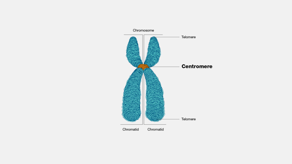
Chromatids
Chromatids refer to the two symmetrical arm-like structures of chromosomes seen during metaphase. During metaphase when the chromosomes are well-defined, each one of them appears to be an elongated body with a matrix and two chromatids.
The chromatids are made of DNA and proteins attached to each other by centromere. They separate during anaphase when sister chromatids migrate to the respective daughter cells.
Chromonema
According to classical cytology, chromosomes are composed of chromonema and centromeres. In less compact chromosomes, under the microscope, a coiled filament is seen in chromosomes. In 1912, Vejdovski named this coiled filament, chromonema.
Chromonema are early-stage chromatids seen as thin filamentous structures during prophase. It is synonymous with chromatid but chromonemata specifies the single molecule of linear DNA with proteins. It also bears genes.
Chromosomes may contain two, four, or more fibers as per the species. The number of threads in chromonema varies with the phases and may have 1-4 threads or fibrils of chromonema. Such coils can be of two types- paranemic and plectonemic.
- Paranemic coils are formed when the chromonema is separated from the coils easily.
- Plectonemic coils are formed when the chromosomal coil stays connected to the coils which makes it difficult to separate.
The degree of coiling in the chromonemal coiling at meiosis and mitosis is variable and often depends on the length of the chromosomes.
Chromomeres
The chromonemata possesses numerous bead-like structures called chromomeres. These bead-like structures are formed by the condensed chromatin material during interphase. A chromatid with chromomeres looks like a beaded necklace. These regions consist of highly coiled or folded DNA.
The portion of the chromomeres in the chromonema is consistent for a given chromosome. The region between the chromomeres is called inter-chromomeres.
Centromere and Kinetochore
During the metaphase and anaphase, the chromosome becomes thicker and takes a filamentous form. At this point, each chromosome will have a clear area known as a centromere or kinetochore. Chromosomes having a centromere are called centric and those without a centromere are called acentric.
- The centromere is the region that is attached to the spindle fiber during cell division.
- They lie in the thinner, primary constriction region of chromosomes.
- This centromere divides the chromosome into two parts or arms. Structurally, the centromere is more light-staining than the other parts, especially during metaphase and anaphase.
- During these phases, there will be a clear zone where the fibrils are uncoiled or less coiled.
- Granules of varying sizes may be attached to these fibrils.
- These are also the same fibrils that constitute chromosomes.
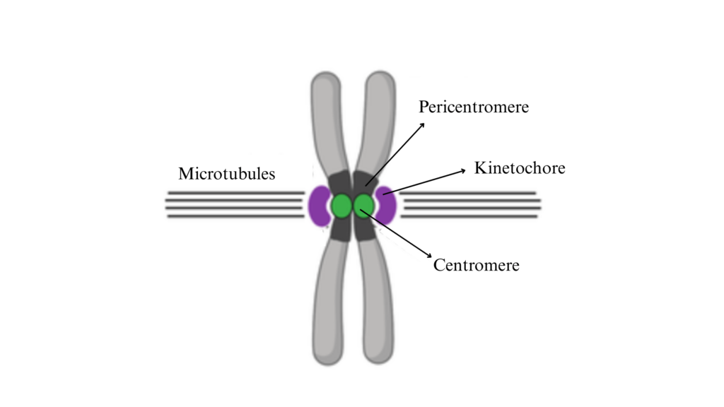
Centromeres have highly repetitive DNA and some specific DNA sequences and proteins to form disc-like structures called kinetochores.
- Kinetochore has a dense outer proteinaceous low-density middle layer and dense inner layer.
- They are bound to centromeres.
- During division, microtubules of spindle fibers attach to the kinetochore and help with chromosome movement during anaphase.
- It serves as an assembly center for polymerization of proteins to form microtubules.
Telomeres
The terminal ends or the extreme ends of chromosomes are called telomeres. It possesses some specific characteristics. They may have a polarity that prevents other segments from fusing to it. Broken ends of chromosomes lack this polarity and can fuse together.
Satellites
Some chromosomes contain a knob-like, elongated, or round appendage called satellites that are connected to the chromosomes via thin chromatin filament. These satellites may have the same diameter as the chromosomes or smaller in size. Generally, at least one chromosome will have a secondary constriction and a satellite attached to it.
Additional Structures
Secondary constrictions in chromosomes are additional structures at any point on chromosomes. These structures help identify the chromosomes as they show deviations or bends.
Nuclear organizers are areas in the secondary constriction that have genes that code for 28s, 18s, and 5.85s rRNA and that induce nucleoli formation.
References
- Agarwal, P. V. |. V. (2004). Cell biology, Genetics, Molecular Biology, Evolution, and Ecology: Evolution and Ecology. S. Chand Publishing.
- https://www.uou.ac.in/sites/default/files/slm/BSCBO-301.pdf
- https://bmellone.uconn.edu/research.html