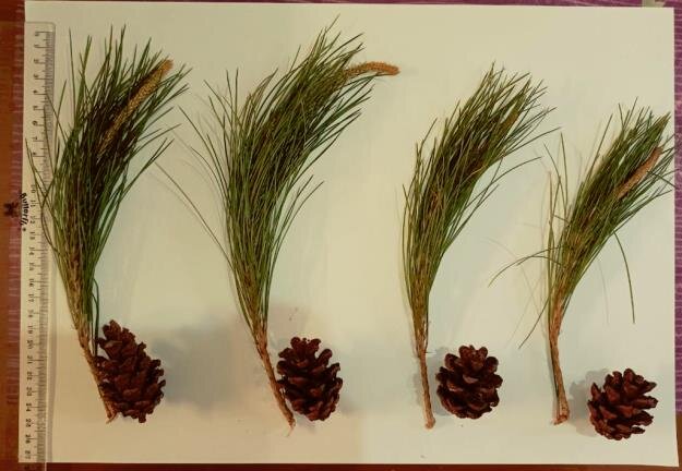
The Pinus plant is dimorphic, with two types of leaves- scale leaves and foliage leaves.
The green foliage leaves make the Pinus plant evergreen. These leaves arise from the spur shoots. They are needle-like, cylindrical, and 20-40 cm long. The anatomy of Pinus needle shows the presence of chlorophyll, which helps it persist for a longer period, making the plant evergreen.
T S of Pinus Needle
The cross-section of the Pinus needle appears circular, semi-circular, or triangular in outline. This shape depends on the species. Despite the difference in outline, the anatomical features of the Pinus needle are the same across all species. The anatomy of the Pinus needle shows its xerophytic characteristics.
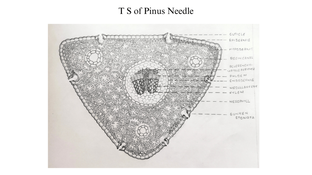
- The epidermis has a thick cuticle layer outside.
- Sunken stomata are seen on the epidermis.
- The stoma opens externally into a cavity called the vestibule and internally into the substomatal cavity.
- Hypodermis contains a few layers of sclerenchyma, with more such cells in the corners.
- Cortical cells underneath sclerenchyma cells are parenchymatous.
- They contain chloroplast and starch grains.
- These parenchyma cells have infoldings in the inner walls and are called arm parenchyma.
- Mesophyll may have resin cells.
- Endodermis follows the mesophyll.
- Pericycle is multilayered, composed of parenchymatous cells.
- These parenchymatous cells store starch.
- Albuminous cells are seen in between the endodermis and phloem, and they serve as channels that transport substances between phloem and mesophyll.
- Pericycle contains tracheid cells as well.
- These cells have bordered pits.
- They are radially elongated and conduct food from the xylem and transport it to the mesophyll.
Albuminous cells and tracheid cells together constitute the conductive tissue between mesophyll and vascular bundles. They compensate for the lack of side veins.
Vascular Bundle in Pinus Needle
The anatomy of Pinus needle shows two vascular bundles in the centre. Some species have only one bundle. The radial longitudinal section of the Pinus needle passes through the xylem, phloem, and cambium showing longitudinal orientation while the medullary rays are seen running horizontally.
- Sclerenchyma cells are seen in between two vascular bundles.
- These cells are unbranched and pass parallelly through the centre of the needle.
- Each vascular bundle has a ventral xylem and dorsal phloem in triangular needles.
- In semi-circular needles, the xylem is towards the flat side and the phloem is located on the curved side.
- The protoxylem is directed to the ventral side and the metaxylem is towards the phloem. Such a vascular bundle is called collateral and endarch.
- Xylem contains tracheids but no vessels.
- The xylem is composed of tracheids with oblique end walls.
- The bordered pits on the radial walls are seen in the surface view.
- In the longitudinal section, these bordered pits show the middle lamella overarched by a dome-shaped area formed by lignified walls.
- They are uniseriate and are separated by crescent-shaped bars of special thickening made of cellular tracheidal walls.
- Phloem has sieve cells and phloem parenchyma.
- The sieve cells are with tapering ends.
- Sieve areas present in radial walls and companion cells are absent.
- The Pinus needle does not have companion cells.
- Medullary rays are uniseriate,
- They encase resin canals and become multiseriate fusiforms.
- Rays in the phloem region consist of starch-filled cells surrounded by albuminous cells.
The xerophytic characteristics seen in the anatomy, help them retain water and make it evergreen, even in winter.
References
- Abraham P C. Bryophytes, Pteridophytes, Gymnosperms & Paleobotany. 2000. St. Mary’s Books & Publications.