The anatomy of Cycas is unique for each part of the plant. The older plants have thick, columnar, and woody stems covered with leaf bases. Here we will see the anatomical features of Cycas stem, leaves, rachis, and roots.
Anatomy of Cycas Stem
A cross-section of the Cycas stem shows an irregular exterior due to the attachment of leaf bases.
- It has a single layer of epidermis having several inferences and leaf traces.
- The cortex is multilayered and is made of parenchyma cells.
- These parenchyma cells contain starch grains.
- This stored starch is the source of sago.
- It also has mucilage ducts and calcium oxalate crystals.
- There are no endodermis or a pericycle.
Stele
- The stele of Cycas has several vascular bundles.
- Vascular bundles are small and appear in the form of a broken ring.
- Each vascular bundle has its phloem on the outer side of the cambium.
- The primary xylem has only tracheids and is endarch.
- Metaxylem shows scalariform thickening.
- Protoxylem has a spiral thickening.
- The cambium is non-distinct and is short-lived.
- A narrow band of parenchyma separated the primary xylem from the primary phloem.
- There is a large pith made of parenchyma cells.
- The pith occupies the centre and contains starch.
- Most often mucilage canals are also seen.
Secondary Thickening in Cycas Stem
Due to the presence of a large amount of parenchyma in between the vascular bundles, the stem of Cycas is very soft and is called manoxylic wood. During secondary growth, which is similar to that in a dicot stem, the cambium produces secondary cells to form a continuous ring.
The cambium that produces the secondary xylem and phloem towards the inside becomes inactive and new cambial cells are formed to continue the secondary growth. This continues several times to form the alternate rings of the xylem and phloem.
Towards the outside, cork cambium produces phellogen and phelloderm that thicken the exterior of the stem. Later on, lenticels are also formed on the Cycas stem.
Leaf Traces
A large number of leaf traces, obliquely pass through the cortex of Cycas stem. The leaf trace emergence and their connection to the base of the periole creates an Omega shape.
- Leaf traces create leaf gaps in the main xylem which causes macrophyllus types.
- Each leaf has 4 leaf traces in pairs, called direct leaf traces and girdle leaf traces.
- Direct Leaf Traces: Two of these leaf traces are formed directly towards the side of the leaf.
- Girdle Leaf Traces: The other two leaf traces are formed opposite to the side of the leaf. They emerge from the stem stele curve in opposite directions and encircle or girdle around the vascular cylinder before entering the leaf.
Both direct and girdle leaf traces arise from the protoxylem of the vascular bundle of the primary ring and then pass through the cortex and enter the leaf.
These leaf traces divide and redivide to form a large number of bundles before entering the rachis. These bundles are usually endarch in stem, pseudomesarch or mesarch when entering the rachis and become exarch at the terminal end of the rachis.
Anatomy of Cycas Root
Internally, the Cycas root resembles that of a dicot root.
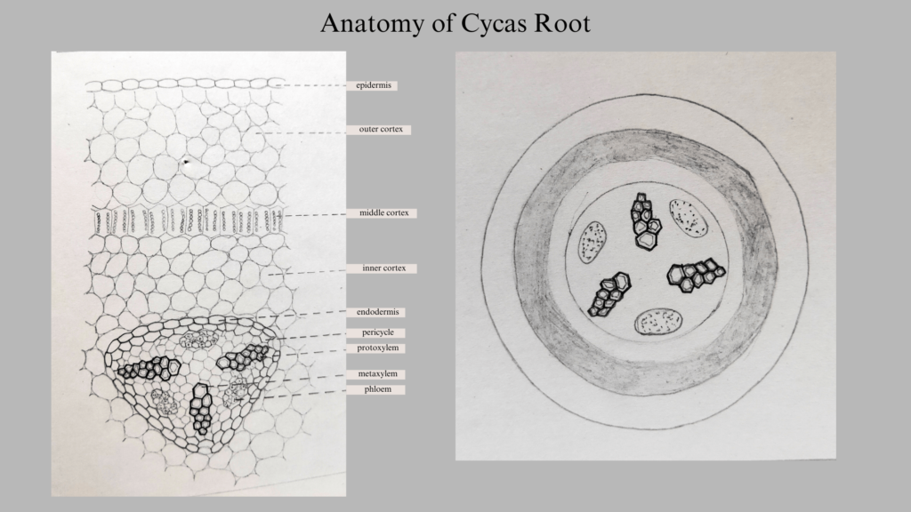
- It has a single-layered epidermis with root hairs.
- A broad parenchymatous cortex follows the epidermis.
- Mucilage canals and cells with tannins are seen in the cortex.
- The endodermis contains Casparian strips, which form the next layer.
- A single-layered pericycle having starch grains comes next.
- The stele contains diarch, triarch, and tetrarch xylems with the phloem located between them. This type of vascular bundle is called radial.
- Protoxylem is seen towards the periphery and this is an exarch xylem.
- Metaxylem has pitted or scalariform thickening.
- Phloem contains phloem parenchyma and sieve tubes.
- Older roots have bast fibers in the phloem.
Secondary Growth in Cycas Root
Secondary growth in the Cycas root occurs in the same manner as in the case of a dicot root. A few parenchyma cells present in between the xylem and phloem, and that surrounding the phloem become meristematic.
- They start functioning as cambium.
- These cambial strips look crescent-shaped.
- The cells of the cambium start cell division to produce the secondary xylem towards the centre and the secondary phloem towards the periphery.
- Broad parenchymatous rays are also formed from the cambium.
- Thus, the cambium produces a manoxylic secondary wood.
- In the meantime, the pericycle behind the protoxylem becomes meristematic and starts functioning as a cambium.
- This will produce more cells to join the crescent ray cambium strips.
- These cells produce living parenchymatous cells opposite to the protoxylem points or the primary medullary rays.
Cork Cambium
The cork cambium of phellogen produces the outermost layer of the cortex below the epidermis. It produces dead cells of phellum towards the outside and living secondary cork cambium or phelloderm towards the inside. These three layers form the periderm. After secondary growth, the epidermis of Cycas root is replaced by periderm. Later, old roots form lenticels.
Anatomy of Rachis of Cycas
The petiole of the compound leaf is called the rachis. The outline of the Cycas rachis appears biconvex or round. The upper lateral sides have depressions coinciding with the leaflet bases.
- The first layer is the cuticle followed by the epidermis.
- Sunken stomata are present in the epidermis.
- The next layer is chlorenchyma cells and a hypodermis formed by sclerenchymatous cells.
- This is followed by parenchymatous ground tissue.
- There are calcium oxalate crystals and mucilage canals in this region.
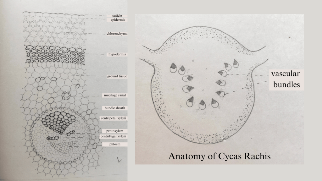
Vascular Bundles
Vascular bundles are embedded in the parenchymatous region.
- Vascular bundles are collateral, conjoint, open, and endarch.
- They are arranged in the shape of Omega.
- Each vascular bundle contains an xylem, phloem, and a strip of inactive xylem.
- The remaining space is occupied by phloem.
- Each bundle has two types of xylem
- An exarch xylem is seen on the upper side of the rachis, appearing as a triangle.
- The metaxylem here is on the upper side and the protoxylem is towards the lower side.
- Two linearly oriented small endarch xylem groups appear on the lower side.
- The two xylem groups are separated by parenchymatous cells.
- In the second group of xylem, the protoxylem is towards the upper side and the metaxylem is towards the lower side.
- Due to the presence of both endarch and exarch, it is called pseudomesarch or diploxylic.
- The phloem is seen below the endarch xylem.
- A strip of inactive cambium is present in between the xylem and phloem.
T S of Leaflet of Cycas
Cycas’s leaflet has a dorsiventral habit. The internal structure shows a cuticle followed by an epidermis on both sides of the leaflet. The epidermis is unilayered and thick. Sunken stomata are seen on the lower epidermis.
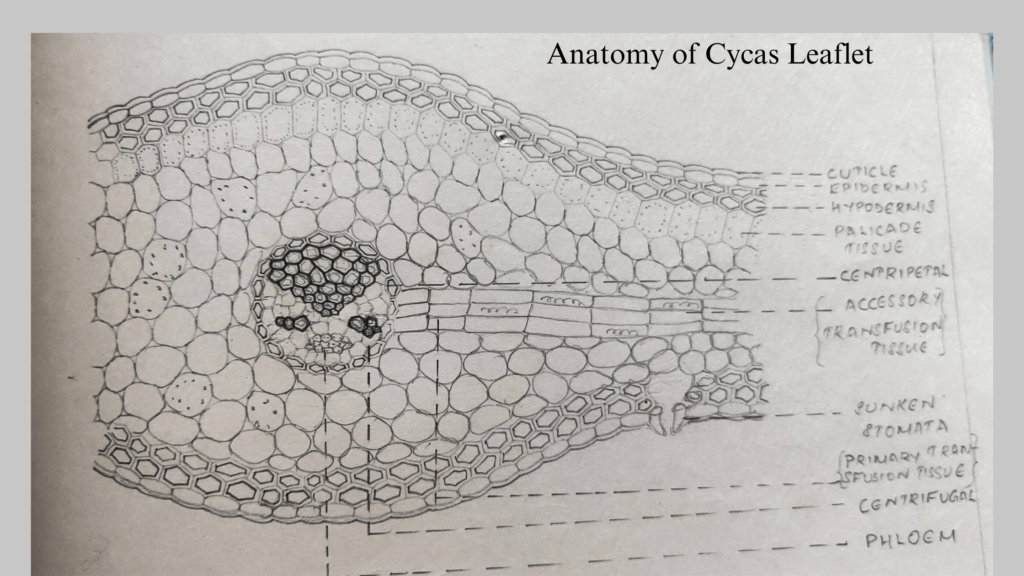
Hypodermis
Hypodermis is sclerenchymatous for protection and mechanical support. Sclerenchyma is absent below the stomata where it opens to the mesophyll.
Mesophyll
- Mesophyll is parenchymatous and is differentiated into upper palisade and lower spongy tissue.
- Palisade has compactly arranged columnar cells with chloroplast.
- Spongy tissue is multilayered containing air pores and chloroplasts.
Vascular Bundle
- The vascular bundle has an endodermis followed by a pericycle.
- The xylem is exarch on the upper side and endarch on the lower side.
- Parenchyma cells separate the xylem and phloem.
Transfusion Tissue
Radially arranged groups of elongated cells arising from the vascular bundle form the transfusion tissue. It passes through the centre of mesophyll. It conducts water and food to the lateral sides of the leaflet. There are primary and accessory transfusion tissues.
Primary transfusion tissue resembles tracheids with bordered pits. They are seen on both the upper and lower sides.
Accessory transfusion tissue has laterally elongated cells arising from the midrib to the sides. They appear in between the palisade and spongy tissue. They help conduct food and water to the lateral sides. This is also considered an extension of the centripetal xylem.
Xerophytic Adaptations of Cycas Leaflet
Cycas plants are found in semi-arid areas and these plants show xerophytic features or adaptations.
- The Cycas leaflets are leathery and tough in texture.
- The epidermis has a thick cuticle.
- Sunken stomata present on epidermis.
- Hypodermal sclerenchyma reduces transpiration.
- Only one vein is present in the leaflet to reduce lateral conduction.
- Spongy mesophyll tissues contain air spaces.
- Air spaces reduce the evaporation and diffusion phases of transpiration.
- Transfusion tissue compensates for side veins.
References
- Abraham P C. Bryophytes, Pteridophytes, Gymnosperms & Paleobotany. 2000. St. Mary’s Books & Publications.
- https://littleflowercollege.edu.in/upload/e_contents/files/eb53e7cabfee73ca13df5254c1f5de74.pdf
wohh just what I was searching for, regards for putting up.